The cardiology department at Valais Hospital treats over 2,000 patients a year. It performs nearly 3,000 cardiac ultrasounds annually and over 1,600 coronary angiograms and coronary angioplasties (stent placements) each year. Among the most frequently performed tests and procedures are:
Examinations and procedures
Cardiac imaging techniques
Echocardiograms
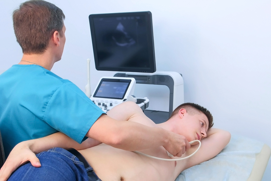
Echocardiograms: techniques that use ultrasound to visualise the heart and its valves. There are several types of cardiac ultrasound: transthoracic, transoesophageal, stress and exercise echocardiograms, and microbubble tests.
Cardiac magnetic resonance imaging (MRI)
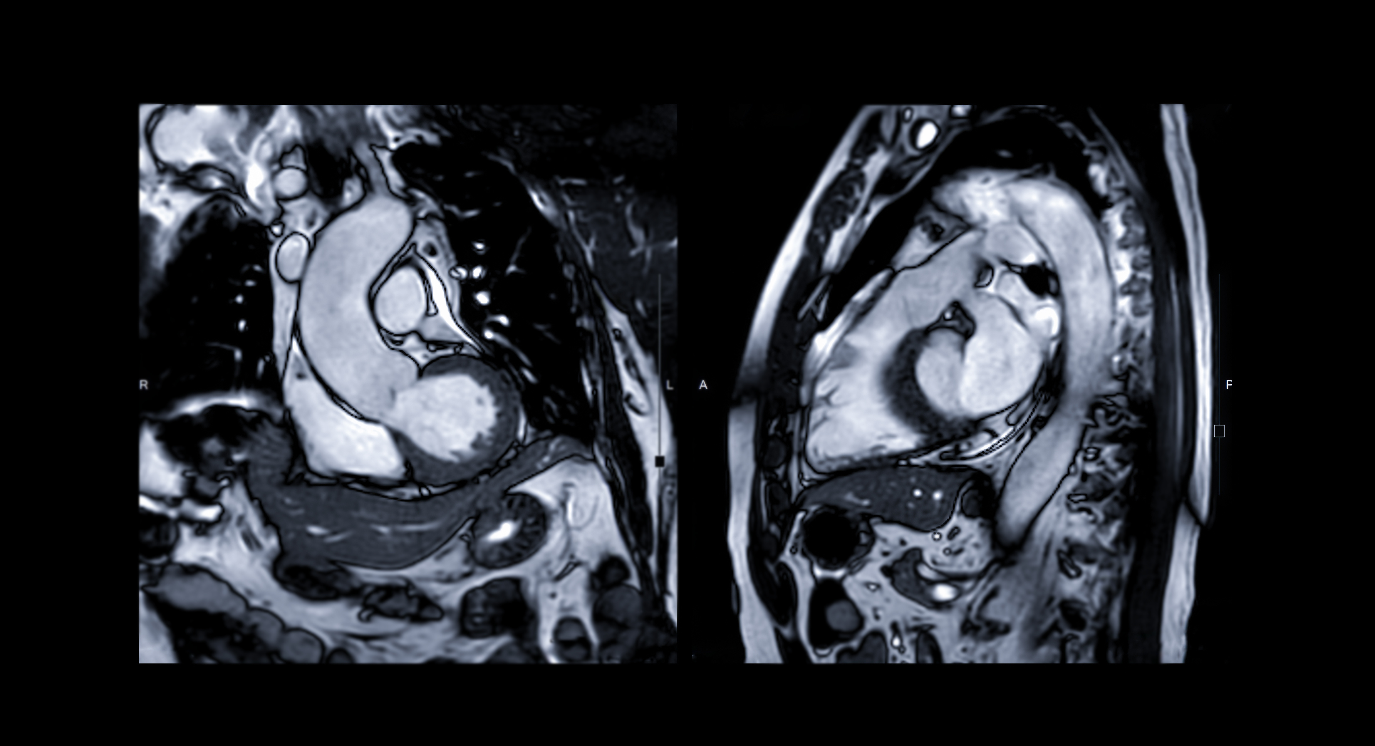
Cardiac magnetic resonance imaging (MRI) and nuclear medicine imaging techniques (myocardial scintigraphy and cardiac PET scans) may be used to supplement the assessment, depending on the individual case.
Interventional cardiology
Interventional cardiology enables the diagnosis and treatment of heart conditions using a percutaneous approach (without opening the chest).
Coronary angiography
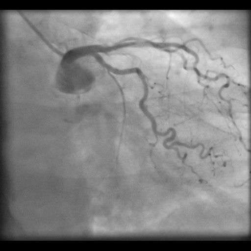
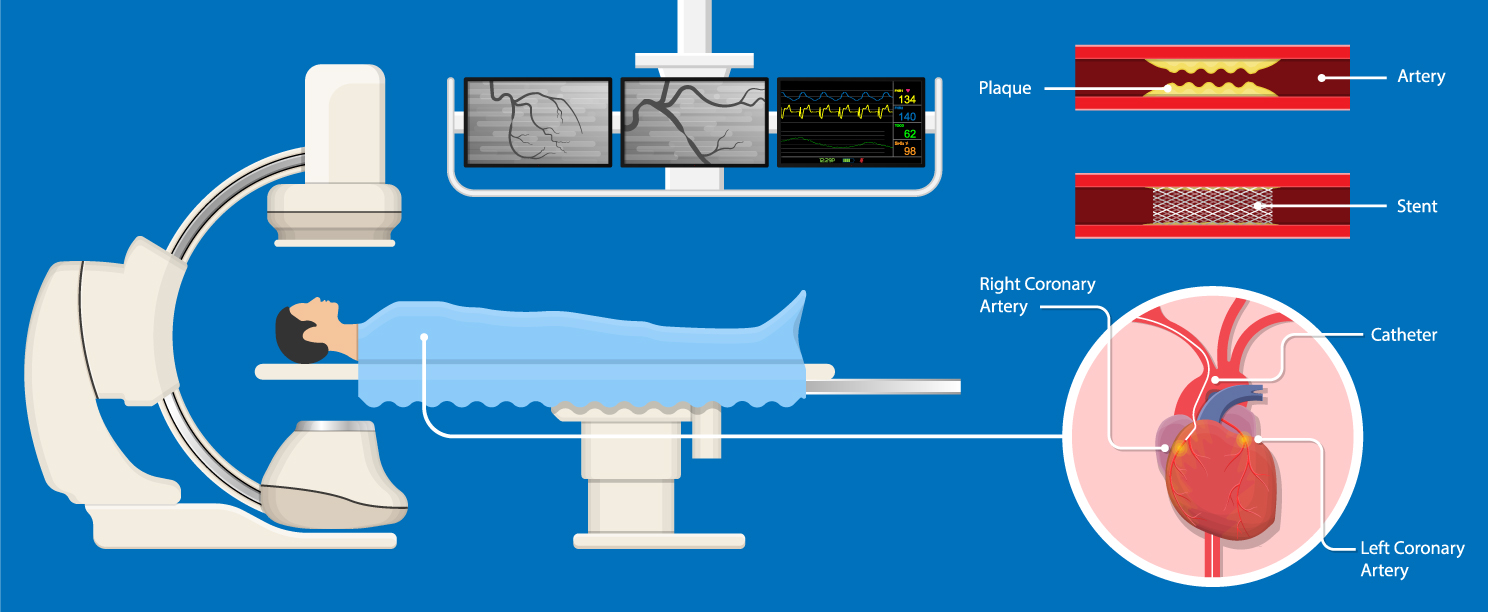
Coronary angiography is an invasive X-ray examination of the heart’s arteries, which may be combined with a study of pressure levels in the heart and lungs known as cardiac catheterisation. A puncture is made in the artery in the thigh (femoral artery) or wrist (radial artery) under local anaesthetic. A catheter (flexible tube) is guided to the opening of the coronary arteries. An iodinated contrast agent is injected and X-rays are taken, enabling any narrowing of the arteries to be diagnosed. This examination is also necessary to screen for damage to the heart’s arteries prior to major surgery, such as an organ transplant, heart valve surgery or surgery on the large blood vessels.
Coronary angioplasty
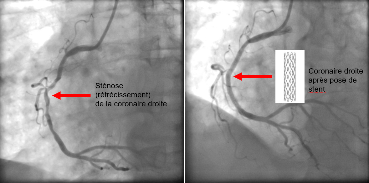
Coronary angioplasty involves inserting a balloon into the narrowed or blocked artery and inflating it to restore sufficient width to the coronary artery. The cardiologist then almost always implants a stent (a small spring-like device), which helps to keep the artery open.
You can watch a video presentation of outpatient interventional cardiology at Sion Hospital here.
Percutaneous repair of congenital heart defects
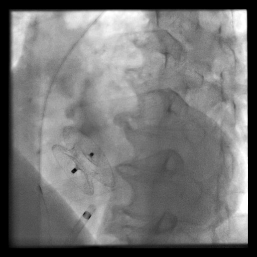
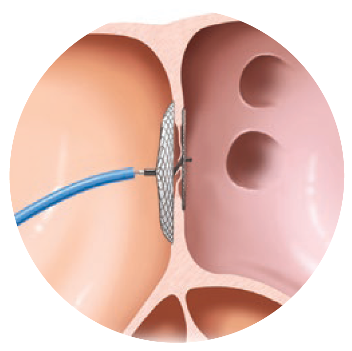
Percutaneous repair of congenital heart defects mainly involves the percutaneous closure of a patent foramen ovale (PFO) or an atrial septal defect (ASD). These are ‘holes’ in the wall between the heart’s atria. After puncturing the right femoral vein under local anaesthetic, a catheter is guided up into the right atrium of the heart. A double-umbrella-shaped device is then deployed across the opening between the atria to seal it.
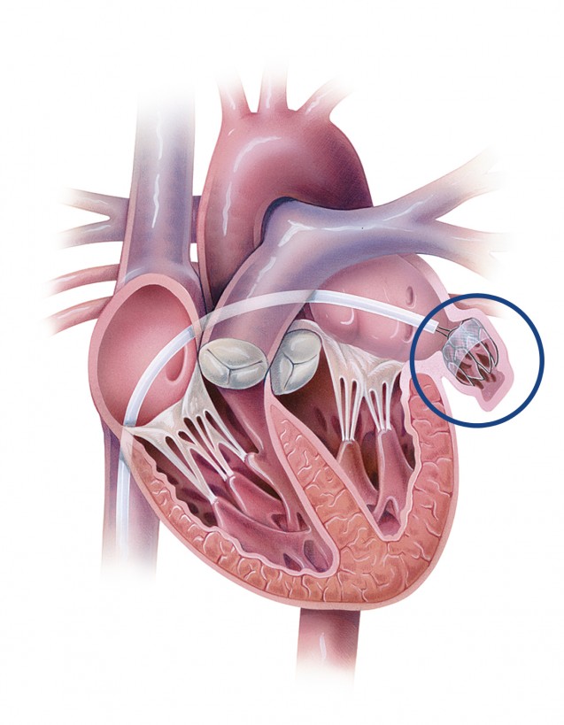
Percutaneous closure of the left atrial appendage
The closure or occlusion of the left atrial appendage involves preventing the formation of a blood clot in patients with atrial fibrillation. However, it is not a first-line treatment. In fact, patients with atrial fibrillation are often prescribed anticoagulant therapy. However, given the risk of haemorrhagic complications, which can sometimes be serious, its use is sometimes contraindicated in certain individuals. It is for these patients that left atrial appendage closure is particularly appropriate.
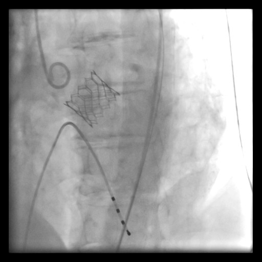
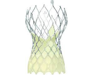
Transcatheter Aortic Valve Implantation (TAVI)
Transcatheter Aortic Valve Implantation (TAVI) is used to treat aortic valve stenosis without the need for traditional heart surgery, particularly where the surgical risk is too high.
Renal artery denervation
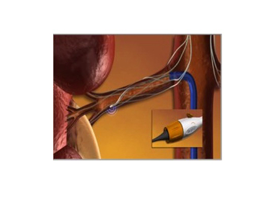
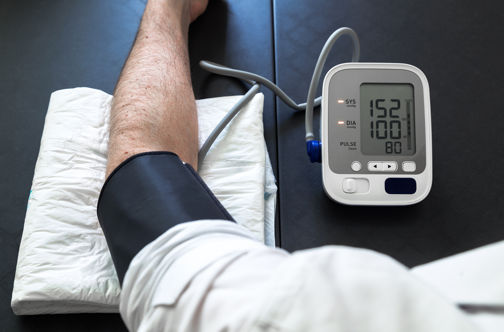
Renal artery denervation involves inserting a radiofrequency probe into the renal arteries and ablating the nerve fibres responsible for refractory hypertension.

Diagnosis and treatment of cardiac arrhythmias
Electrophysiological study (EPS)
The electrophysiological study (EPS) aims to use catheters (long, thin tubes usually inserted into a vein in the thigh (femoral vein)) to document heart rhythm disorders. Fluoroscopy and magnetic fields are used to guide the catheters inside the heart. These catheters enable the electrical activity of the heart to be recorded and electrical conduction tests to be carried out.
La thermo ablation ou ablation par radiofréquence
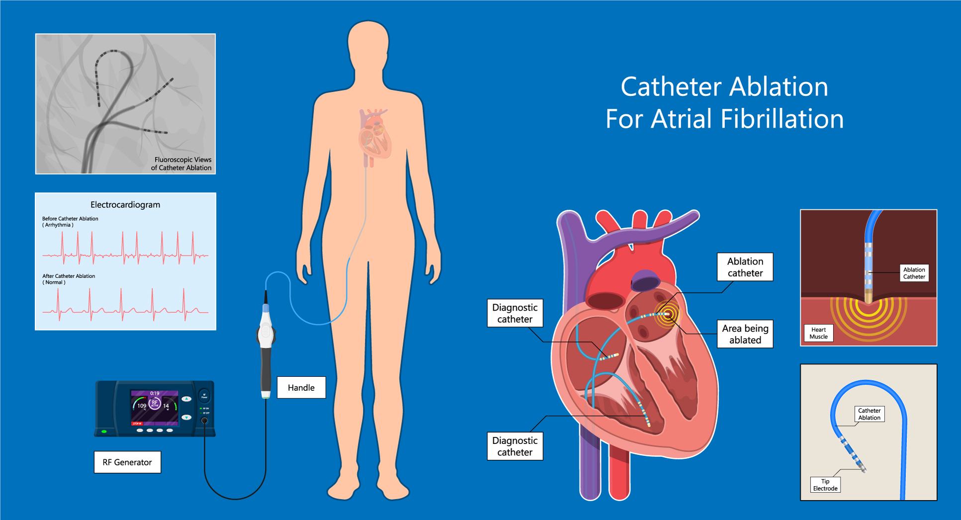
La thermo ablation ou ablation par radiofréquence permet de supprimer par l’application d’un courant électrique la zone responsable du déclenchement des arythmies, notamment flutter, fibrillation auriculaire et tachycardie ventriculaire.
The implantation of a pacemaker and a defibrillator
![[Translate to English:] Stimulateur cardiaque (pace maker)](/fileadmin//images/disciplines/cardiologie/AdobeStock_210372386-_1_.jpg "[Translate to English:] Stimulateur cardiaque (pace maker)")
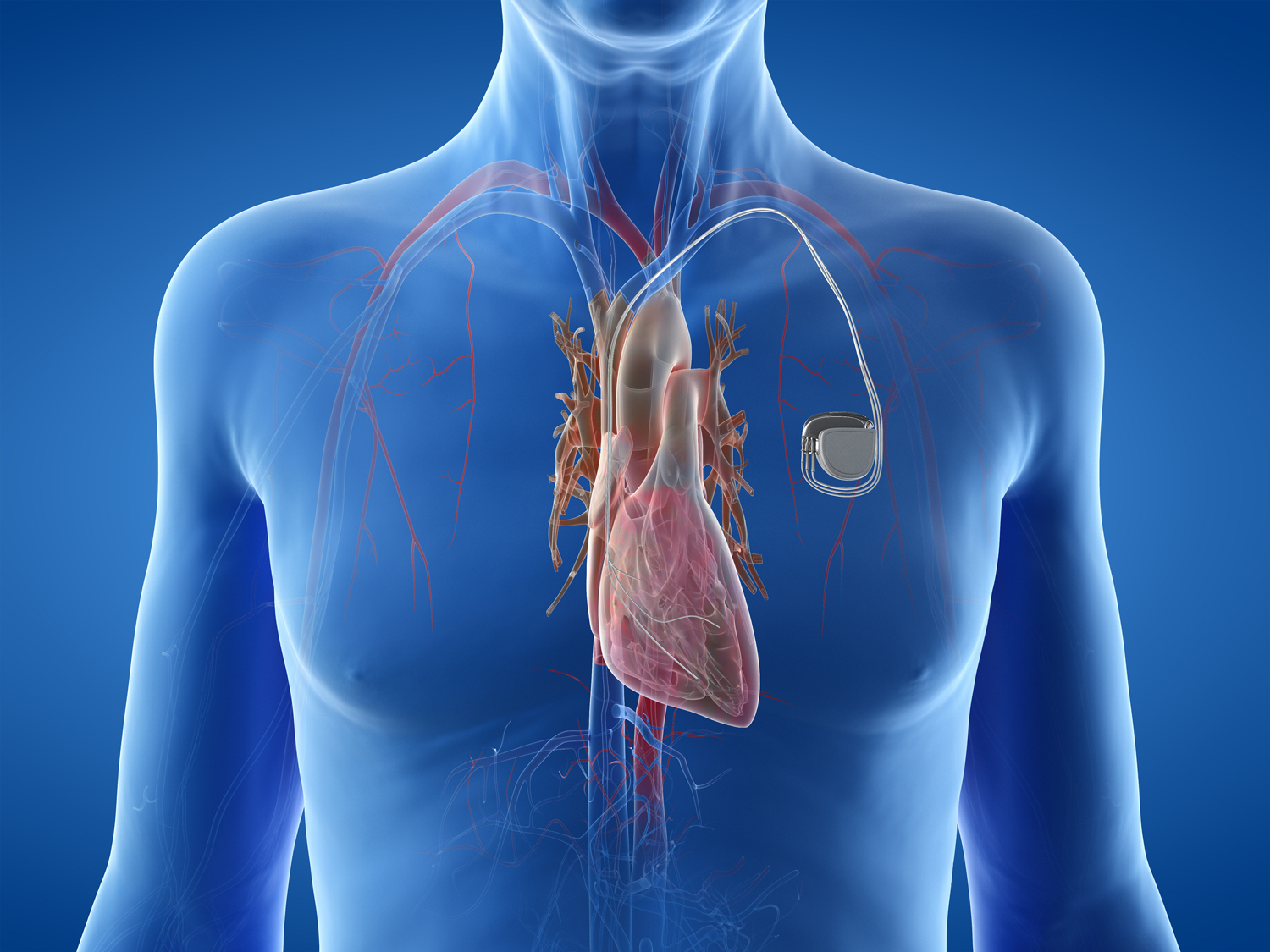
The implantation of a pacemaker and a defibrillator, or the replacement of a pacemaker’s housing, is carried out under local anaesthetic by a cardiologist or a cardiac surgeon. An incision of a few centimetres is made under the collarbone, after which the surgeon inserts one or two electrodes via a vein beneath the collarbone; these are then secured to the heart muscle. In some cases, a third electrode may be inserted to stimulate the left ventricle and perform cardiac resynchronisation. The defibrillator is implanted during a procedure similar to that for a pacemaker. In addition to its function as a pacemaker, the defibrillator is capable of interrupting an abnormally rapid heart rhythm, which could lead to loss of consciousness or even sudden death.
Source vidéos: © Medtronic
Les maladies cardiaques
Notre équipe de cardiologie est formée de cardiologues FMH expérimentés dans la prise en charge des différentes affections cardiaques, qu’elles concernent les artères du cœur, les vaisseaux, le muscle cardiaque, les valves ou les troubles électriques.
Vous trouverez des informations relatives aux différentes affections sur le site de la fondation suisse de cardiologie.
Voici une liste des principales pathologies que nous pennons en charge :
La vidéo suivante résume la prise en charge en cas d’insuffisance cardiaque.
La vidéo suivante résume la prise en charge en cas d’infarctus du myocarde.
Brochures et informations
- Information pour les patients avant cathétérisme cardiaque et coronarographie
- Conduite à tenir en cas d’urgence
- Insuffisance cardiaque - Vos médicaments
- L’athérosclérose - Les facteurs de risque
- La Nitroglycérine
- Traitement du rétrécissement de la valve aortique par voie percutanée (TAVI)
- Suivi ambulatoire de l’insuffisance cardiaque
- Chirurgie cardiaque
- Centre ambulatoire de réadaptation cardiaque et vasculaire : informations aux patients
- 20 ans chirurgie cardiaque et de la cardiologie en Valais